X-rays are a primary tool for early identification of dental problems. Detecting issues with x-rays before they become problematic can save you money in the long run. Early detection can help prevent the need for more extensive, expensive procedures or surgeries. X-rays are primarily used to detect:
• Internal tooth decay
• Cysts (fluid filled sacks at the base of your teeth)
• Tumors, both cancerous and non-cancerous
• Impacted teeth
• Teeth that are still coming in
Digital X-Rays
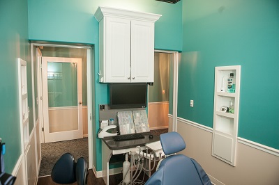
At Cane Bay Family Dentistry, we use digital x-rays, which have several advantages over traditional film based x-rays. Digital x-rays allow us to take x-rays with 1/5 of the radiation that you would receive from traditional dental x-rays. The worry of exposure to excess radiation is eliminated.
Large on-screen x-rays make patient communication more effective. The immediate observation of the images on the screen allows us to discuss your dental health quickly and accurately.
Digital radiography has greatly enhanced the practice of dentistry. It allows the patient and the doctor to see images of the teeth in higher resolution on a large format for easier detection of problems, all while decreasing the radiation exposure to the patient. The process we use further defines the radiograph, resulting in clinically meaningful images that are sharp, detailed and rich in contrast.
Images are available instantly after exposure, eliminating the wait and effort spent developing and mounting x-rays. If an image needs to be retaken, it can be done immediately. Digital format also allows us to send and receive your images electronically, allowing for a faster consultation with your dentist.
Ultrasonic Scaler
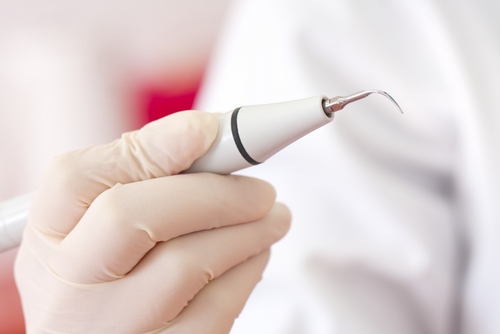
We use ultrasonic scalers for most adult dental cleanings. These devices use ultrasonic vibrations to help break down the plaque and calculus on the teeth that cause gingivitis and periodontal disease. The ultrasonic cleaners create microscopic bubbles that implode on the surface of the tooth, killing microbes and removing plaque and tartar in the process. Our instruments use a thin tip to better navigate in the periodontal pockets to help maintain optimal gingival health.
The procedure uses water and/or an antimicrobial liquid called chlorhexidine. It can remove tartar buildup in hard-to-reach areas, with no damage to the tooth enamel. Manual scaling often uses pressure for cleaning, while the vibration produced by the scaling tip of an ultrasonic scaler is barely perceptible. This makes ultrasonic cleaning suitable to those with sensitive teeth. The cleaning process is faster than manual scaling, making your visit more comfortable.
After your teeth have been cleaned with the ultrasonic cleaner, your teeth will be hand scaled to check for any residual deposits and then polished.
Intraoral Camera
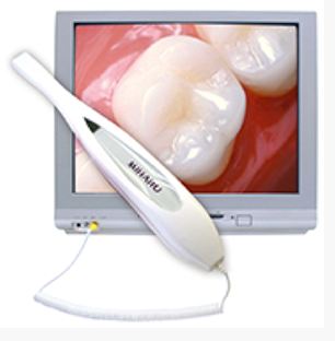
X-rays give us a clear view of what we can’t see such as decay between the teeth. An intraoral camera gives the patient a view of what the dentist sees. An intraoral camera is a device that is about the size of a toothbrush. Broken fillings, fractured teeth, decay or any other dental problem can be viewed so that we can discuss with you any treatment needed. With a clear understanding of your dental needs we can decide together what the best treatment is for you.
CBCT
At Cane Bay Family Dentistry, we utilize the CBCT system to take 3D images of your teeth. 3D imaging allows us to better understand the anatomy of your mouth, which is essential before we perform any type of corrective procedure. By using this state-of-the-art technology, our team will be better prepared to accurately diagnose potential issues and develop the best possible treatment program for you.
A typical dental x-ray will just focus on the teeth, and for each image you’ll need one exposure. Therefore, to get the same picture as a 3D image, you’d need many exposures. 3D imaging shows considerably more than a simple 2D x-ray, as this newer technology will provide more accurate and complete visual information from every angle. Additionally, the data can be easily shared and duplicated without the worry of film getting lost.
Best of all, the system allows us to properly diagnose your oral and facial impairment while exposing you to minimal radiation levels. Our 3D scanner will allow us to select the perfect scanning area or field of view, helping to limit your exposure to radiation since we will be focusing directly on the area of concern.
After the scan, the information will be used to develop a proper treatment plan for your oral or facial issue. We’ll use this technology to easily and quickly share images of the problem area with your referring dentist or physician. This will allow your doctors to work together on your care to deliver a positive treatment experience.
PATIENT TESTIMONIALS
Our patients have wonderful things to say about the care they received from our practice:
